You know how some diseases just sound like they belong in a sci-fi movie? Well, Caroli disease is one of those. It’s like the universe decided to throw a curveball at our liver’s bile ducts, causing them to go all crazy and dilated. Not fun, right?
So, picture this: you’re living your life, maybe enjoying a slice of pizza (or two). Then boom! You find out you have this rare condition. Talk about a plot twist! It’s not just about the symptoms; it’s about figuring out what to do next.
Radiology comes into play here, shining a light on what’s happening inside your body. It helps doctors see the extent of the disease and plan how to manage it best. Sounds pretty important, huh?
In this little chat we’ll unpack how these imaging insights can make a real difference in handling Caroli disease. Buckle up!
Advancements in Radiological Imaging Techniques for Diagnosing Caroli Disease: A Comprehensive Review
Caroli disease is this rare genetic condition that affects the liver, leading to the dilation of bile ducts. And because it’s not super common, diagnosing it can be a bit tricky. But thanks to some cool advancements in radiological imaging techniques, things are looking up!
First off, let’s talk about **ultrasound (US)**. It’s often the first step doctors take when they suspect something might be wrong with your liver. A simple, non-invasive procedure that uses sound waves to create an image of your organs. In Caroli disease, the ultrasound can show those enlarged bile ducts quite clearly. You know how sometimes you see a distorted reflection in a funhouse mirror? That’s kind of what happens here; it highlights those dilated ducts that are characteristic of the condition.
Then you’ve got **computed tomography (CT)** scans. CTs offer a more detailed view than ultrasounds. They take multiple x-ray images from different angles and put them together to create cross-sectional views of your body—a bit like slicing a loaf of bread and looking at each piece! For Caroli disease, CT scans can help spot complications like infections or even biliary stones that might form because the bile ducts aren’t working right.
Now let’s not forget about **magnetic resonance imaging (MRI)** and particularly a technique called MR cholangiopancreatography (MRCP). Think of MRCP as an advanced version of MRI tailored for looking at your bile ducts and pancreas specifically. It gives super detailed images without exposing you to radiation, which is always nice! For diagnosing Caroli disease, MRCP shines by allowing doctors to visualize those dilated bile ducts with stunning clarity.
What’s even cooler is how medical professionals are blending these technologies. Sometimes they’ll use multiple imaging techniques for better accuracy. Like when you’re trying to figure out where your friends are at a concert—they’re sending texts from all over! By combining ultrasound data with MRI results, doctors get a fuller picture that might just lead to faster diagnosis and treatment decisions.
But what about after diagnosis? Well, advancements don’t stop there! Tools like elastography—where you measure liver stiffness—can help gauge whether there’s any progression in liver damage caused by Caroli disease. If you’ve ever poked a marshmallow and felt it squish down? That’s kind of like elastography; stiffer liver tissue means trouble!
In summary, advancements in radiological imaging for diagnosing Caroli disease are making strides with:
- Ultrasound: Quick and easy first-step imaging.
- CT Scans: Detailed cross-sectional views.
- MRI/MRCP: High-resolution images without radiation exposure.
- Combining Techniques: Using various methods for higher accuracy.
- Elastography: Assessing liver stiffness post-diagnosis.
These innovations truly play a pivotal role in improving outcomes for patients battling Caroli disease. When diagnosed swiftly and accurately through these methods, individuals can get appropriate treatment sooner rather than later! And that’s something everyone should feel good about—a world where science meets compassion in healthcare.
Comprehensive Overview of Caroli’s Disease Types: Radiological Insights and Imaging Techniques in Medical Science
Caroli’s disease is not the most common condition, but it’s super interesting and a bit complex. It’s basically a genetic disorder affecting your liver’s bile ducts. If you picture your liver like a team of workers, these bile ducts are like the delivery guys taking bile from the liver to the intestines. When they don’t work right, things can get pretty messy.
Types of Caroli’s Disease
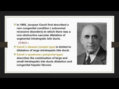
There are two main types: simple Caroli’s disease and Caroli syndrome.
- Simple Caroli’s Disease: This is when you have cystic dilatation of the bile ducts without any other major issues. It’s mostly just those pesky bile ducts getting all swollen up, which can lead to problems like pain or infections.
- Caroli Syndrome: This one’s trickier because it comes with other complications like kidney issues or even congenital heart defects. It’s a combo package that makes it more serious.
So, how do we actually spot these issues? That’s where radiological insights come in.
Imaging Techniques
Doctors rely on imaging techniques to diagnose and manage Caroli’s disease effectively. Here are a few key ones:
- Ultrasound: One of the first tools in the toolkit! It’s non-invasive and uses sound waves to create images of your organs. It can show cysts and structural changes in the liver pretty well.
- CT Scans: A computed tomography scan gives a much clearer view than an ultrasound. It helps identify complications that might arise from those dilated bile ducts, like infections or abscesses.
- MRI: Magnetic resonance imaging is another powerhouse in this field. It’s especially good at providing detailed images of soft tissues, making it easier to visualize complications associated with Caroli syndrome.
- EUS (Endoscopic Ultrasound): This one is super fascinating! It combines endoscopy with ultrasound technology to get even closer to those tricky structures inside your abdomen.
Let’s say you’re having some pain in your upper abdomen and visit a doctor. They might start with an ultrasound just to see what’s going on inside there without any invasive procedures. If they find something odd—like those enlarged bile ducts—they’ll probably follow up with a CT scan or MRI for more details.
Treatment Implications
Once diagnosed, treatment can vary widely depending on which type of Caroli’s disease you have and whether there are other complications involved.
- If it’s simple Caroli’s disease, doctors may keep an eye on things unless you develop symptoms that require action.
- If it’s Caroli syndrome with kidney issues, then it’s a more extensive management scenario due to its complexity—sometimes even involving surgery if necessary.
What really hits home for me about this condition is how much imaging has transformed lives! A friend once shared their experience of dealing with unexplained stomach pains for years until finally—thanks to MRI—they found out what was happening: cysts from Caroli’s disease were causing all those problems!
So yeah, understanding the types of Caroli’s disease along with smart imaging techniques can make all the difference in diagnosing and managing this condition effectively. You follow me? Each tool has its purpose; it’s about finding what works best for each situation!
Advancements in Caroli Disease Treatment: A Comprehensive Overview of Current Scientific Approaches
Caroli disease, huh? It’s one of those rare conditions impacting the bile ducts, making them all weird and enlarged. So, let’s chat about the latest advancements in treating this condition—there’s been some interesting stuff happening lately.
First off, diagnosis is crucial. Radiological insights play a huge part here. Using imaging techniques like MRI and CT scans, doctors can visualize those dilated bile ducts. This helps not only in diagnosing Caroli disease but also in understanding its severity. Picture this: you’ve got a high-def camera showing everything inside your body. It’s kind of mind-blowing!
Then comes treatment options. One common approach involves managing complications rather than the disease itself—like you know, treating infections or addressing liver issues that might pop up. But here’s where things get exciting! There’s been a shift towards more interventional procedures. Doctors are looking into ways to support those bile ducts directly.
For instance, there’s a technique called endoscopic retrograde cholangiopancreatography (ERCP). Sounds fancy, right? Well, during this procedure, docs can actually visualize and treat blockages in the bile ducts. This can relieve symptoms and improve quality of life for patients.
Not stopping there! Some researchers are investigating more innovative methods like surgical interventions. In severe cases where complications are significant, they might recommend surgery to remove parts of the affected bile duct or even perform a liver transplant if it comes to that. It’s not just about cutting stuff out but also ensuring that whatever’s left functions properly.
Another cool area is genetic research. Scientists have started to dive deep into what causes Caroli disease on a genetic level. Some folks believe certain genes can contribute to how the disease develops or progresses. If they nail down these genetic factors, who knows? They might even find ways to prevent it or manage it better long-term.
Lastly, keep an eye on clinical trials! There are always new treatments being tested out there—some may involve medications designed specifically for Caroli syndrome symptoms or complications. It’s almost like being part of a sci-fi movie where new solutions are popping up all around us!
So yeah, Caroli disease treatment is evolving thanks to advancements in radiology and therapy techniques. The future looks bright for those affected by it! Let’s continue hoping for more breakthroughs because every step forward means better lives for patients dealing with this challenging condition.
So, Caroli disease, huh? It’s one of those conditions that doesn’t get enough airtime. You know what I mean? It’s not something you hear people chatting about over coffee. But it’s important because it affects the bile ducts and can lead to some serious health issues.
I remember chatting with a friend who was grappling with this diagnosis. She was stressed and overwhelmed, trying to navigate a sea of medical jargon and treatment options. Honestly, hearing her talk about it really opened my eyes to how crucial understanding this condition is—not just for those directly affected but for the medical community as well.
Now, radiological insights play a massive role in managing Caroli disease. We’re talking MRI scans and ultrasound imaging here—they can reveal changes in the bile ducts that might not be visible otherwise. It’s like looking at a treasure map; without the right tools or perspective, you might miss important landmarks that give clues on what’s happening beneath the surface.
These imaging techniques can help doctors identify complications early on, which is super critical because timely intervention can really change the game for patients. Think about it: when you catch something before it spirals out of control, there’s a better chance of managing symptoms and improving quality of life.
But here’s where it gets tricky. Not every imaging technique offers the same level of detail or clarity. The interpretation is an art as much as it is science—doctors need to read those images with precision and context in mind. It’s like being a detective piecing together clues; one wrong assumption could lead to mismanagement of the condition.
And let’s not forget about patient comfort! While these tests are essential, they can also be intimidating or uncomfortable for some folks. A little empathy and clear communication from healthcare providers can go a long way in easing anxiety.
In short, while radiological insights into Caroli disease management are vital—offering clarity on what can seem like murky waters—it’s equally important to approach treatment holistically. Patients need support beyond just images; they deserve compassion, guidance, and reassurance along their journey. So yeah, when we think about healthcare innovations—it’s all interconnected!