You know what’s wild? The human eye is like this super complex camera, but without the fancy lenses. Seriously! It can do all this amazing stuff, yet we take it for granted every day.
Picture this: you’re out on a sunny day, and bam! Your eyes adjust like magic. Or when you spot that cute puppy from across the street—your eyes zoom in faster than your brain can process it.
Crazy, right? Each part plays its role like an orchestra in a concert, working together to help us see the world clearly. So, let’s break it down with a labeled diagram that makes sense of all these tiny parts and their big jobs. Ready to take a peek at the eye’s secrets?
Exploring the Structure and Function of the Human Eye: A Comprehensive Guide to Ophthalmic Anatomy and Physiology
Sure! Let’s take a stroll through the fascinating world of the human eye. You might not think about it often, but your eyes are like little cameras capturing the world around you. So, what’s going on behind those pretty peepers? Let’s break down the structure and function of the human eye.
The Structure of the Eye
First off, let’s chat about the major parts of the eye. Each section plays its own unique role in helping you see everything from a beautiful sunset to a friend’s smile. Here are some key components:
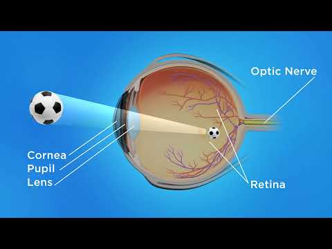
- Cornea: This is like your eye’s window. It’s clear and helps focus light onto the retina.
- Iris: The colored part of your eye! It controls how much light enters by adjusting the size of the pupil.
- Pupil: The black circle in the center of your iris. It changes size based on how bright or dark it is outside.
- Lens: Located behind the pupil, it fine-tunes focus for objects that are near or far away.
- Retina: This is where magic happens! Light hits this layer at the back of your eye and turns into electrical signals.
- Optic Nerve: This is like a messenger that sends those signals to your brain, letting you see!
Now you have an idea about which parts we’re working with!
The Functioning of Your Eye
But let’s get into how all these pieces work together to help you see… it’s kind of mind-blowing! When light enters through the cornea, it bends slightly (this process is called refraction). Then, it passes through your pupil and lens, which further refines that focus.
Imagine you’re at a concert and trying to read a sign across the stadium—your lens helps adjust to make that blurry sign crisp and clear. How cool is that?
Once light reaches your retina, photoreceptor cells (called rods and cones) get involved. Rods are super sensitive to light and help you see in dim conditions while cones catch colors in bright light. These cells transform light into electrical impulses.
And here comes a little emotional nugget: Picture being at a family gathering where laughter fills the room, and everyone is sharing stories—those moments captured by our eyes are made possible through this whole system working seamlessly!
Your Brain Takes Over
Once those electric signals travel through your optic nerve to your brain’s visual cortex, they become images! Your brain takes that info from both eyes (that means both perspectives) and combines them to create depth perception. That’s how you can tell if something is close or far away.
So think about when you’re riding a bike; knowing just how far away that parked car really is makes all the difference between smoothly zooming past or becoming one with it!
A Final Thought
Your eyes do so much more than just allow you to see—they connect you with memories, emotions, and experiences every single day. From watching sunsets to reading books late at night or even sharing laughs with friends—everything relies on this incredible structure and function.
The next time you’re taking in something beautiful—or even just looking at your phone—take a moment to appreciate how complicated yet seamless our eyes really are! Seriously, it’s nothing short of amazing!
So there ya go! Now you’ve got an idea about how our vision works. Pretty neat stuff happening behind those eyeballs!
Anatomy of the Human Eye: A Detailed Diagram and Scientific Insights
The human eye is a marvel of biological engineering. When you think about it, this little organ not only lets us see the world but also helps us navigate our everyday lives. Let’s break down its anatomy in a way that’s super easy to digest.
First off, we gotta talk about the basic structure. The eye is basically like a camera, capturing light and sending images to our brain. Here are the key parts you’ll want to know:
- Cornea: This is the transparent front layer of the eye. It bends light so it can enter through the pupil.
- Iris: The colorful part that surrounds the pupil. It controls how much light gets in by adjusting the size of the pupil.
- Pupil: The opening in the center of the iris. It gets bigger or smaller depending on lighting conditions.
- Lens: This clear structure behind the pupil focuses light onto the retina, allowing you to see things clearly up close or far away.
- Retina: A layer at the back of your eye that has special cells called photoreceptors which convert light into electrical signals.
- Optic nerve: Think of this as your eye’s communication line to your brain. It transmits those electrical signals for processing.
So, here’s what happens when you see something pretty cool—like a puppy, or a sunset (cue emotional response!). Light bounces off that object, enters your cornea, passes through your pupil and lens, and then hits your retina. Photoreceptors there (rods and cones) translate that light into signals for your optic nerve.
But wait! There’s more. Did you know that rods are super sensitive to dim light? That means they help you see when it’s dark—like when you’re sneaking into the kitchen for a midnight snack! On the flip side, cones are all about color and detail; they work best in bright light.
Here’s an interesting tidbit: sometimes people have issues with their eyes because these components don’t work quite as they should. For example, if someone has a problem with their lens—say cataracts—they might find their vision cloudy or blurry.
The anatomy doesn’t just stop at these parts either; there are tiny muscles around your iris known as sphincter muscles that help control those smooth movements to adjust lighting without you even thinking about it!
To wrap this up nicely: if you picture all these parts working together like a finely-tuned orchestra, you’ll appreciate just how important each piece is for giving us our beautiful view of life around us every day.
So next time you’re looking at something amazing—a beautiful sunset or that goofy pet—you can think about all those incredible structures doing their job to bring it all into view!
Detailed Anatomy of the Human Eye: Labeled Image for Scientific Study
The human eye is like an incredible camera that captures light and translates it into what we see. It’s super complex, and each part plays a crucial role in how we perceive the world around us. Let’s break it down a little.
Cornea: This is the clear, dome-shaped front surface of your eye. It helps focus light that comes in, acting almost like a lens. Imagine it as the window to your eye! Without a properly functioning cornea, things can look blurry.
Iris: Ever seen someone with striking blue or brown eyes? That’s your iris! It’s the colored part of your eye and controls how much light enters by adjusting the size of the pupil. So when you’re in a dim room, your iris opens wide to let in more light.
Pupil: Right in the center of the iris is the pupil. Think of it like an adjustable aperture on a camera. Its size changes based on lighting conditions; larger in low light and smaller when it’s bright outside.
Lens: Behind the pupil lies the lens. This clear structure fine-tunes focusing by changing shape—thinner for distant objects and thicker for close-ups. It’s kind of like how you zoom in or out on your phone camera!
Retina: At the back of your eye is the retina, which acts like film in traditional cameras. It contains millions of cells called photoreceptors (rods and cones) that convert light into signals sent to your brain. Rods are great for low-light vision, while cones pick up colors.
Optic Nerve: Once those photoreceptor cells do their magic, they send messages through the optic nerve to your brain where everything gets processed into what you see—like sending an email with all those photos!
Sclera: The sclera is basically the white outer layer of your eyeball. It’s tough and protects all those delicate inner structures; think of it as a case for a prized possession.
Aqueous Humor and Vitreous Humor: These are fluids inside your eye that help maintain its shape and provide nutrients to its inner parts. Aqueous humor is found in front of the lens while vitreous humor fills up space behind it.
Understanding all these parts gives us insight into just how amazing our eyes really are! The next time you marvel at a sunset or read a book under soft lighting, just remember—all those intricate components are working together seamlessly so you can enjoy every detail!
So when talking about a labeled diagram, it’s basically used by scientists to illustrate these parts clearly—making studying anatomy way easier, especially if you’re diving into biology courses or even medical studies someday.
Each label helps pinpoint where each piece is located within this tiny universe that resides right inside our heads! Who knew such complexity could fit snugly within something so small?
So, you know when you look at a diagram of the human eye, and it’s all labeled with fancy terms like cornea, retina, and iris? It might seem a bit overwhelming at first, but honestly, these diagrams are super helpful if you want to understand how our eyes work.
I remember the first time I came across one of those diagrams in school. It was a science class, and our teacher pointed to each part like we were on some kind of eye anatomy adventure. I was both intrigued and confused—like how did that little lens help me see my friend across the room? It’s wild to think about!
The eye is a pretty complex organ, right? It captures light and transforms that into signals for our brain to turn into images. Just think about it for a second: all those tiny parts have to work together perfectly every time you blink! The cornea is basically the shield—it protects everything else inside. Then there’s the pupil, which opens and closes depending on how much light comes in. The iris gives it that beautiful color—like mine is brown but my sister’s is green!
And don’t get me started on the retina! That’s where all the magic happens. It collects light rays and turns them into signals that your brain interprets as images. Can you even imagine what life would be like if one of those parts didn’t function correctly?
Anyway, looking at labeled diagrams helps break it down into manageable bits. You can almost feel like you’re piecing together a puzzle about how we perceive the world around us. And honestly, having a visual aid really solidifies your understanding—it connects everything in your mind.
So next time you see one of those diagrams, don’t just glaze over it! Take a moment to appreciate all those beautifully labeled parts working together in harmony so you can watch that sunset or see your best friend smile from across the room. It’s kind of amazing when you think about it!