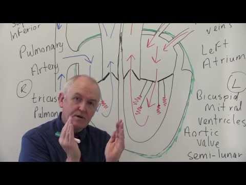
You know what’s wild? The heart, that little fist-sized muscle in your chest, pumps about 2,000 gallons of blood every day! That’s like filling a swimming pool in a week. Crazy, right?
I remember once, I was so fascinated by how it all works that I tried to explain it to my dog. Spoiler alert: He didn’t get it. But the excitement was real!
So, let’s break down the anatomy of this amazing organ. It’s not just a pump; it’s a whole system working behind the scenes. Understanding how it all fits together is like piecing together a puzzle — and trust me, it’s pretty cool stuff!
Comprehensive Guide to Heart Anatomy: Downloadable PDF Resource for Science Students
The heart is an incredible organ, wouldn’t you agree? It’s not just a symbol of love; it’s also a complex structure that plays a crucial role in keeping us alive. Understanding the anatomy of the heart can feel overwhelming at first, but it’s pretty fascinating once you break it down.
First off, let’s look at what the heart actually is. It’s a muscular organ about the size of your fist and sits slightly left of your chest. That muscle, called myocardium, pumps blood throughout your entire body. Pretty vital stuff, right?
Now, let’s talk about its main parts:
- Atria: There are two upper chambers called the left and right atria. They receive blood coming into the heart.
- Ventricles: Below the atria are two lower chambers: the left and right ventricles. These guys pump blood out of the heart.
- Valves: The heart has four valves—tricuspid, pulmonary, mitral, and aortic—that ensure blood flows in just one direction. Think of them like doorways that only allow passage one way.
- Septum: This is a wall that separates the left and right sides of your heart. It keeps oxygen-rich blood separate from oxygen-poor blood.
When we look at how all these pieces work together, it gets even more exciting! Blood flows through this system in two loops: one for oxygen-poor blood to go to your lungs (where it gets fresh oxygen) and another for oxygen-rich blood to supply your body.
Speaking of blood flow—let’s break down its journey a bit more:
1. Blood enters through the superior and inferior vena cavae into the **right atrium**.
2. It moves through the tricuspid valve into the **right ventricle**.
3. From there, it travels through the pulmonary valve to get pumped into the lungs via pulmonary arteries.
4. Once it’s picked up oxygen in the lungs, it returns via pulmonary veins to enter the **left atrium**.
5. Next up is passing through the mitral valve into the **left ventricle** which then sends that fresh oxygen-rich blood out through the aortic valve into your body.
Can you imagine all this happening every second? It’s like an endless cycle!
Now if you’re studying this stuff—maybe in class or just out of curiosity—you might find diagrams super helpful! These visuals can really show you where everything goes inside that awesome little pump we call our heart.
Oh! And remember when I mentioned how important those valves are? If they don’t work well, it can lead to some serious health issues like regurgitation or stenosis. Knowing where these structures are located helps in understanding such conditions better.
So yeah—the anatomy of our hearts isn’t just academic; it’s simply amazing! If you’re looking for more resources or diagrams to aid your studies—like downloadable PDFs or comprehensive diagrams—there’s plenty out there made just for science students like you.
Just think about all that beats away inside us—it makes you feel grateful for every heartbeat!
Comprehensive Overview of Heart Anatomy and Physiology: Downloadable PDF Guide for Science Enthusiasts
Sure! The heart is like the superstar of our body, pumping blood and keeping everything running smoothly. Knowing how it’s built and how it works can be super cool. Let’s break it down a bit.
Heart Anatomy
Your heart is basically a muscular pump, about the size of your fist. It’s divided into four main chambers:
- Right Atrium: This is where deoxygenated blood enters from the body.
- Right Ventricle: It pumps that deoxygenated blood to the lungs for oxygenation.
- Left Atrium: Oxygen-rich blood returns from the lungs here.
- Left Ventricle: This powerful chamber pumps oxygenated blood out to the rest of your body.
You know, just thinking about my own heart makes me remember this one time when I was so nervous before giving a speech in front of class. My heart was racing like it was in a marathon! That’s just how it reacts to stress sometimes.
Now, let’s get into those little details that make your heart function so well.
Valves
Your heart has valves that act like doors, controlling the flow of blood:
- Atrioventricular Valves: These include the tricuspid valve (between right atrium and ventricle) and the mitral valve (between left atrium and ventricle).
- Semi-lunar Valves: These are found at the exit of each ventricle: pulmonary valve leads to the lungs, while aortic valve leads to the body.
These valves open and close with every heartbeat. If they didn’t work right, oh boy! Blood would flow backward, which isn’t good news for anyone.
The Heart’s Electrical System
Ever heard about how your heart has its own rhythm? That’s thanks to its electrical system!
- Sinoatrial (SA) Node: Often called the “natural pacemaker,” this tiny area in the right atrium sends electrical signals that trigger each heartbeat.
- Atrioventricular (AV) Node: This node catches signals from SA Node and slows them down before they hit the ventricles. Think of it as traffic control!
If you’ve ever felt your heart skip a beat or race after exercise, now you know why!
Cardiac Cycle
The cardiac cycle describes all events during one heartbeat. Here are two key phases:
- Systole: When your heart contracts and pumps blood out.
- Diastole: When your heart relaxes and fills up with blood again.
This constant cycle keeps everything flowing smoothly through your body—like being on an endless ride on a roller coaster!
So yeah, understanding all these details about heart anatomy and physiology gives you insight into why we feel what we feel during different activities—exercise makes it pump harder; emotions can make it race!
In case you’re interested in visuals or diagrams, try looking up comprehensive diagrams that illustrate all these parts together; they really help bring everything to life!
Keep exploring, because there’s always more to learn about how our incredible bodies work!
Detailed Diagram of Heart Structure: Understanding Cardiovascular Anatomy in Science
Alright, let’s get into the heart of the matter—literally! The human heart is a pretty amazing pump that keeps your blood flowing. It’s not just a simple muscle; it has different parts with their own jobs, working together like a well-oiled machine. So, if we break it down, we can really appreciate how neat and complex it is.
The heart has four main chambers: two atria and two ventricles. Think of them as rooms in a house where blood gets sorted out. The atria (upper chambers) are like entryways. They receive blood from the body and lungs. The right atrium gets deoxygenated blood (that’s blood without oxygen) from the body, while the left atrium takes in oxygen-rich blood that comes from the lungs.
Now, once the atria fill up with blood, they contract and push that blood down into the ventricles. The right ventricle then sends that deoxygenated blood to the lungs for fresh oxygen through the pulmonary arteries. Meanwhile, the left ventricle—this one’s pretty powerful—pumps oxygenated blood out to your whole body through the aorta. It’s kind of like sending packages to different places!
- Aorta: Largest artery in your body; it carries oxygen-rich blood away from your heart.
- Pulmonary veins: These bring oxygen-rich blood back from your lungs to the left atrium.
- Pulmonary arteries: They take deoxygenated blood from the right ventricle to your lungs.
- Cava veins: Superior and inferior vena cava return deoxygenated blood to the right atrium.
You also can’t forget about valves! They are crucial for keeping everything flowing in one direction. There are four valves: tricuspid valve, pulmonary valve, aortic valve, and mitral valve. Each one opens and closes at just the right moment so that there aren’t any traffic jams inside your heart!
I remember when I first learned about this stuff back in school. It was during a biology class where our teacher brought in an actual model of a heart—like wow! Everyone got super involved, poking around at all these parts. It made such a difference seeing it hands-on rather than just reading about it in books.
The walls of each chamber also have different thicknesses. The ventricles have thicker walls than the atria because they need more muscle power to pump that blood out into circulation. Just think about how hard your heart works every day; it’s no joke!
If you want to appreciate what’s going on inside you even more, check out diagrams online or even real-life models if you can find them—it’s fascinating stuff! Understanding this cardiovascular anatomy helps us grasp why keeping our hearts healthy is super important for our overall well-being.
The intricate design really shows nature’s engineering skills at work—you know? It’s all interconnected, working seamlessly together every single second of our lives!
You know, hearts are pretty amazing little systems. They don’t just pump blood; they’re like the central hub of our bodies. I remember the first time I really looked at a heart anatomy diagram in school. There were all these colorful labels and arrows pointing here and there. At first, it kinda felt overwhelming—like trying to solve a puzzle without knowing what it looks like! But once I started to understand things like the atria and ventricles, it all clicked together.
So let’s break it down a bit. The heart is basically made up of four main chambers: two upper ones called atria and two lower ones called ventricles. The right side takes in deoxygenated blood and sends it to the lungs to get more oxygen—think of it as handing off dirty laundry for a fresh wash! Meanwhile, the left side pumps that freshly cleaned blood out to the rest of your body.
And those valves? Totally crucial too! They help keep everything flowing in one direction so you don’t end up with a traffic jam in your veins. It’s wild how something so small can have such an intricate setup, right? When I think about how this little guy keeps our whole system running smoothly, I can’t help but feel grateful for my own heart working away day in and day out.
When you start studying heart anatomy diagrams, you begin to appreciate just how complex yet beautifully designed this organ is. It reminds me that before there were high-tech medical technologies, people had no choice but to study the anatomy closely—like real-life detectives piecing together clues without any fancy gadgets!
So yeah, whether you’re looking at diagrams for class or just out of curiosity, each line and label tells a part of an incredible story about life itself. And honestly? That’s pretty awesome if you ask me!