You know that moment when you’re sitting around with friends and someone suddenly goes pale, clutching their stomach? Yeah, I’ve been there too. You start wondering if it’s just a bad burrito or something more serious.
Well, appendicitis is one of those sneaky culprits, right? One minute you’re fine, the next you’re doubled over in pain. It’s wild how quickly things can change! Just think about it—appendicitis is the classic “surprise” in the world of health emergencies.
What’s even crazier is how radiology has stepped up its game to catch this sneaky appendicitis. We’re talking about some seriously cool tech that helps doctors figure out what’s going on inside your belly without you having to go under the knife first.
So, let’s dig into these advances in radiology. I promise it won’t be boring; there’s some pretty fascinating stuff happening that could help save a lot of people from unnecessary surgeries! Cool, right?
Understanding the Gold Standard for Diagnosing Appendicitis: A Comprehensive Analysis in Medical Science
So, let’s chat about appendicitis and how doctors figure out if you’ve got it. You know, the whole “my stomach hurts” thing can be really confusing. Appendicitis is basically an inflammation of your appendix—a tiny tube sticking out from your large intestine. When it gets angry and swollen, it can cause some serious pain, especially down in your lower right abdomen.
Now, diagnosing appendicitis has evolved a lot over the years. Back in the day, doctors mostly relied on physical exams and patient symptoms to make their call. But as you might guess, that wasn’t always super accurate. So, here comes the **gold standard**: imaging tests.
When we talk about advances in radiology for diagnosing appendicitis, two main tools come into play: **ultrasound** and **CT scans**.
Ultrasound is often used first in kids or pregnant women, since it avoids radiation exposure. It uses sound waves to create images of internal organs. It’s non-invasive and pretty quick—like snapping a picture of what’s happening inside you! However, sometimes it’s hard to see everything clearly.
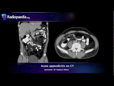
Then there’s the CT scan, which has become like the superhero of appendicitis diagnosis. Basically, it gives a much clearer picture because it combines multiple X-ray images taken from different angles and turns them into a detailed cross-sectional view of the abdomen. Doctors can spot inflammation around the appendix or even any complications like abscesses with this method.
But here’s where things get interesting! You might wonder how doctors choose one method over another? Well:
- Patient age: Like I said earlier, kids and pregnant women usually get ultrasounds first.
- Gender: In women of childbearing age, physicians may consider gynecological issues too.
- Symptoms: If someone shows classic signs—like pain migrating from around the belly button to the lower right—they might quickly move on to a CT scan.
- Access to technology: Some places just don’t have fancy CT machines everywhere.
The sensitivity of these methods is important too! That means how good they are at actually spotting appendicitis when it’s there—not missing it or waving off something serious as nothing. CT scans are known for high sensitivity levels compared to ultrasounds.
However—here’s something crucial—you still wanna balance accuracy with potential radiation risks from repeated CT scans over time. Doctors weigh those factors carefully while deciding what route to take.
Interestingly enough, there are also new techniques on the horizon! For example:
- MRI scans: They’re being tested and could come handy since they don’t use radiation either!
- Laparoscopy: Sometimes doctors go straight in for a look if they think surgery might be needed anyway.
One final thought: trust your gut—literally! If you’re feeling off or experiencing severe abdominal pain along with nausea or fever? Don’t hesitate; just go see someone who knows what they’re doing!
So yeah, that’s sort of where we’re at with diagnosing appendicitis today: a mix of history and cutting-edge tech that helps save time and lives by catching problems early on!
Understanding the Puylaert Technique: A Key Method in Medical Imaging Science
The Puylaert technique is a pretty cool method used in medical imaging, especially for diagnosing appendicitis. You might be wondering, what’s so special about it? Well, let’s break it down a bit.
First off, this technique uses **ultrasound** to spot appendicitis. It’s one of those nifty tools that doesn’t rely on radiation, which is great since too much exposure to X-rays can cause problems down the line. So, you know, it’s like having a safe way to peek inside your body without all the risks!
So how does this work? The Puylaert technique involves placing the patient in a specific position—usually lying on their side with their knees drawn up. This helps shift the intestines and makes it easier for doctors to get a clear view of the appendix. When done right, the ultrasound can reveal if the appendix is inflamed or even if there’s an abscess nearby.
Now, here are some key points about why this technique shines:
I remember when my friend Sarah had to go through this whole process. She was feeling all sorts of pain in her belly and was anxious about what was going on. Thankfully, her doctor used the Puylaert technique. The ultrasound showed that her appendix was indeed inflamed, and she got treated without wasting any time!
Something important to mention—while the Puylaert technique is super helpful for many cases of appendicitis, it does have its limits. For instance, if someone’s appendix is positioned unusually or if they have other conditions mimicking appendicitis symptoms (like ovarian issues in women), things can get tricky.
In recent years, advances in radiology have really boosted how we diagnose appendicitis with techniques like this one becoming more common in emergency settings. Plus, more training for doctors ensures they’re using these methods effectively.
To sum up: the Puylaert technique has made a significant mark in medical imaging science. By combining safety with efficiency, it helps doctors detect issues quickly while minimizing risks associated with radiation exposure. So next time you hear about someone getting an ultrasound for belly pain—know that this clever method could be at play!
Evaluating Appendicitis in Pregnancy: Preferred Imaging Modalities and Their Clinical Implications
Evaluating appendicitis in pregnancy can be quite tricky. You’ve got a whole bunch of factors at play here, and that adds some serious complexity to the diagnosis. So let’s break it down, shall we?
First off, appendicitis can be harder to diagnose in pregnant women because their bodies go through so many changes. As the uterus enlarges, it can push everything around. Pain that you might think is due to appendicitis could actually be from pregnancy-related issues. This makes imaging super important.
When it comes to imaging modalities, there are a couple of preferred methods for diagnosing appendicitis during pregnancy:
- Ultrasound (US): This is usually the first-line choice. It’s safe for both mom and baby since it doesn’t use radiation. Doctors often start with an ultrasound to look for signs of appendicitis by checking for fluid or inflammation.
- Magnetic Resonance Imaging (MRI): If ultrasound results are inconclusive, MRI comes into play. It’s another radiation-free option that’s pretty effective at visualizing abdominal organs and can help confirm an appendicitis diagnosis.
- Computed Tomography (CT): While CT scans are great at spotting appendicitis, they involve radiation—which isn’t ideal during pregnancy. That said, if a doctor really suspects appendicitis and other methods aren’t cutting it, they might consider using a low-dose CT.
So, what’s the deal with these imaging methods? Here’s where clinical implications get interesting. Early diagnosis is key when dealing with appendicitis in pregnant women because delayed treatment can lead to complications like perforation or even sepsis—yikes!
Let me share a brief story: A friend of mine experienced severe abdominal pain while pregnant. Initially misdiagnosed as just pregnancy cramps, she eventually had an ultrasound that revealed her appendix was indeed inflamed. Luckily, they caught it in time! But this whole situation highlights how crucial accurate imaging can be.
In summary, evaluating appendicitis in pregnant women primarily involves ultrasound and MRI due to their safety profiles. CT scans are typically reserved for cases where it’s absolutely necessary but come with risks that need careful consideration.
Keeping these things straight is vital for healthcare providers so they can make informed decisions when diagnosing this potentially serious condition!
So, let’s talk about radiology and, specifically, how it’s changed the game for diagnosing something like appendicitis. Honestly, I still remember that time my friend was in so much pain. We thought it was just a stomach bug at first. But then the doctor suggested an ultrasound, and bam! It turned out to be appendicitis. Crazy how quickly things can change with just a little imaging technology.
Basically, when you think of appendicitis, it’s often that dreaded pain in your lower right abdomen. But pinpointing it can be tricky because other stuff can feel similar—gastroenteritis or even kidney stones. This is where rad folks come in with their machines!
You know how they used to just rely on physical exams and patient history? Well now there are these fancy techniques like ultrasound and CT scans that make things way more precise. An ultrasound is super useful for kids or pregnant women since it doesn’t involve radiation—huge plus, right? You get those sound waves capturing images without the scary stuff.
On the flip side, if you’re looking for detailed slices of your insides, a CT scan can provide some pretty vivid images of your appendix and surrounding areas. It helps doctors see if there’s swelling or maybe even abscesses forming. I mean, it’s kind of wild when you think about how we can look inside our bodies without surgery!
Radiology isn’t just about finding problems; it’s also about avoiding unnecessary procedures. Sometimes doctors used to jump straight to surgery after a diagnosis based on symptoms alone. Now they can have a better idea before taking someone to the operating room which not only saves time but also reduces risks for patients.
And hey—imagine being in college back in the day when all this tech was still evolving; dealing with these intense pains while hoping you didn’t have to go under the knife! Thanks to advances in imaging techniques today, people are getting diagnosed faster and hopefully feeling less anxious as they wait for answers.
In short, radiology has made diagnosing appendicitis more accurate and less daunting than ever before. It’s fascinating how technology keeps improving our understanding of health issues around us every day!