You know that moment when you’re sitting in a doctor’s office, and they start throwing around words like “sacroiliitis”? Yeah, me too. Honestly, it sounds like a character from a sci-fi movie or something!
Well, sacroiliitis is no joke. It’s that annoying inflammation in your sacroiliac joints—where your spine meets your pelvis. If you’ve ever felt some gnarly pain in your lower back or hips, it might just be trying to tell you something.
Now, the cool part? Radiology has made some serious strides lately. It’s like upgrading from a flip phone to the latest smartphone—you can do so much more! New tech means doctors can spot these pesky issues faster and with way better precision.
So hang tight! We’re diving into some pretty amazing advances in radiology for diagnosing sacroiliitis. You’ll be surprised at how far we’ve come in figuring this stuff out!
Exploring Innovative Treatments for Sacroiliitis: Advances in Pain Management and Inflammation Control
Sacroiliitis can be a real pain in the back—literally! It’s when the sacroiliac joint, which connects your lower spine and pelvis, gets inflamed. And if you’ve ever had to deal with that kind of discomfort, you know it can seriously impact your daily life.
So, what’s new on the horizon for treating this pesky condition? Well, there’s been some exciting progress lately in how we manage pain and inflammation.
Recent Advances in Treatment
One big step forward has been the use of biologics. These are medications made from living organisms that help target inflammation more specifically. They work by blocking certain proteins that contribute to inflammatory processes. It’s like having a sniper go after the bad guys instead of using a shotgun approach.
Another treatment gaining traction is radiofrequency ablation. It sounds fancy, but it’s basically using heat to disrupt the nerves carrying pain signals from the joint. Imagine turning down the volume on an annoying radio; you still have music, but it’s not blaring in your ears anymore.
Also, physical therapy isn’t just an old-school option; it’s evolving too! Innovative techniques like dry needling and manual therapy are being integrated into treatment plans to help ease tight muscles around the joint. This can lead to better mobility and less pain over time.
Diving into Radiology Advancements
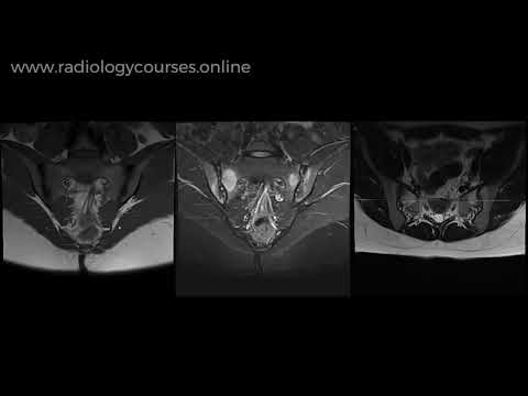
Now onto something techy: radiology. Recently, doctors have improved how they diagnose sacroiliitis through advanced imaging techniques like MRI and ultrasound. These methods not only diagnose issues earlier but also help monitor how well treatments are working over time. Picture it as a high-definition camera capturing everything going on inside without having to go under the knife.
When you’re diagnosed sooner or more accurately with these advanced tools, it’s easier for your doctor to tailor a treatment plan just for you. This personalized approach can make all the difference between managing pain effectively or grappling with chronic issues.
Things to Keep in Mind
While innovations are great—like shiny new toys—they don’t come without their challenges. You might experience side effects from medications or need multiple sessions of physical therapy before feeling better. So patience is key!
Moreover, not every treatment works for everyone. Some folks find relief with biologics while others might swear by physical therapy alone. It really depends on individual circumstances—what works wonders for one might not do much for another.
And hey, always keep communication open with your healthcare provider! They’ll guide you through options based on what’s best suited for your unique situation.
In summary: Sacroiliitis doesn’t have to dictate how you live your life anymore with all these innovative treatments popping up alongside advances in diagnostic technology. With a bit of hope and some work together with medical professionals, managing this condition is becoming more effective than ever before!
Understanding the Gold Standard Test for Sacroiliac Joint Pain: A Comprehensive Overview in Orthopedic Science
Sacroiliac joint pain can really mess with your day, right? It’s that discomfort you feel in your lower back or buttocks that seems to come out of nowhere. This joint, located where your spine meets your pelvis, is a big deal because it bears a lot of weight and helps with movement. So when something goes wrong there, you want to get a clear picture of what’s happening.
Now, let’s talk about the gold standard test for diagnosing issues with the sacroiliac joint. The name sounds fancy, but basically, it refers to the most reliable way doctors figure out if that joint is causing your pain. This involves a mix of clinical evaluation and imaging techniques. The clinical evaluation usually starts with a good chat—your doctor will ask about your symptoms and medical history to get an idea of what might be wrong.
Once they have some background info, they often perform a few tests to see how your body moves and responds. You might have some tenderness in the area or experience pain when certain movements are done. These physical tests can give valuable clues about whether the sacroiliac joint is at fault.
But here’s where it gets interesting: imaging techniques play a huge role here too! Advances in radiology have changed the game for diagnosing conditions like sacroiliitis (that’s inflammation around the sacroiliac joint).
Radiologists often use modalities such as:
- X-rays: They’re like those old-school photos of bones but can’t show everything.
- MRI: This one uses magnets and radio waves to create detailed images. Super helpful for spotting inflammation!
- CT scans: They provide cross-sectional images and can identify structural issues.
These imaging studies can help visualize any changes in bone structure or soft tissue around the joint. So when doctors combine the physical exams with these advanced imaging tools, they get a pretty good view of what’s going on.
However, there’s still some debate about how much weight should be given to these imaging results versus clinical findings. Sometimes you might see changes on an MRI but not feel any pain, which complicates things further! It’s kind of like being caught between a rock and hard place—you want answers, but sometimes those answers aren’t as clear-cut as you’d hope.
So imagine this: you go into the clinic feeling awful because every step sends shooting pain through your back. The doctor runs through their normal routine—chatting about how you’ve been feeling—and then decides an MRI would help clarify what’s up. As you wait for those results, you’re crossing fingers hoping it’s nothing major!
You know what I mean? Getting diagnosed is not always straightforward; it feels like putting together a puzzle without having all the pieces yet.
In short, understanding **sacroiliac joint pain** involves more than just asking “what hurts.” You’ve got this mix of clinical evaluations and cutting-edge radiology working together to give doctors better insight into what might be going on inside your body.
When it comes down to it, everyone wants relief from their pain as soon as possible—but knowing how each method contributes can make that search for answers feel less daunting!
Optimal Diagnostic Approaches for Sacroiliitis: A Comprehensive Review of Scientific Investigations
Sacroiliitis, you know, is inflammation in the sacroiliac joints, which connect your lower spine to your pelvis. It can cause serious discomfort and pain in your lower back and hips. So, nailing down a diagnosis is pretty important for treatment, right?
First off, let’s talk about some of the diagnostic approaches out there. Doctors usually start with a good ol’ physical exam. They’ll poke around to see where it hurts—classic stuff! But that alone isn’t enough since many conditions can mimic sacroiliitis.
Next up is imaging techniques. You might hear about X-rays first. They’re great for spotting bone changes but often miss early signs of inflammation. That’s where MRI comes into play—super useful because it can show inflammation even if there aren’t clear changes in bones yet. Seriously, an MRI is like having a sneak peek into what’s going on under the surface.
Another approach that’s gaining traction is using ultrasound. It’s less common than MRI but still gets the job done sometimes. Ultrasound can help visualize fluid accumulation or other inflammatory markers directly at the joint.
Now let’s break down these options a little more:
- X-rays: Good for rule-outs; they show bony abnormalities but not early soft tissue issues.
- MRI: The gold standard! It reveals inflammation in both bone marrow and soft tissue.
- CT scans: Useful for detailed anatomical views but not as good as MRI for soft tissue issues.
- Ultrasound: Quick and portable; good for seeing fluid or swelling around joints.
And look, all these methods have their strengths and weaknesses. A study showed that while MRIs are awesome for spotting **active sacroiliitis**, they might not be readily available everywhere—like if you’re in a small town without fancy hospitals.
In addition to these imaging techniques, blood tests can supplement the diagnosis. They check for levels of certain markers like C-reactive protein or HLA-B27 antigen that may indicate inflammatory conditions.
But here’s the kicker: diagnosing sacroiliitis isn’t always straightforward! Sometimes doctors have to use a combination of these methods to get it right—or they might even need to follow up with repeat imaging over time to see how things progress.
All in all, what you really want is an approach that tailors the diagnostic process to each patient’s situation—considering their symptoms, medical history, and lifestyle factors too! It’s about piecing together puzzles in unique ways so that people get effective treatments tailored just for them instead of one-size-fits-all solutions.
So yeah, that’s basically what goes down when diagnosing sacroiliitis today! A mix of traditional exams combined with advanced imaging technologies makes sure we’re catching things as early as possible so folks can get back to living life without pain—which is totally what we want!
So, let’s talk about sacroiliitis and how radiology has stepped up its game in diagnosing it. I mean, just saying “sacroiliitis” sounds kinda intimidating, right? It’s this inflammation of the sacroiliac joint, which usually sits at the bottom of your spine where it connects to the pelvis. People with this condition often experience lower back pain, and let me tell you, that can really disrupt your life.
A couple of years ago, my buddy Jake was complaining about his back hurting like crazy. He thought it was just a result of all those late-night gaming sessions! But after a few months, he decided to see a doctor. That’s when the whole radiology thing popped up. They recommended an MRI as part of his diagnosis process. Initially, he felt nervous about the machine—who wouldn’t? But once he was in there, he realized it wasn’t so bad after all!
MRI has become such a powerful tool in diagnosing sacroiliitis because it gives super detailed images of soft tissues and bones. It reveals inflammation better than x-rays do; those tend to miss subtle changes early on. Because sacroiliitis can sometimes be tricky to spot at first glance—like that one friend who always hides behind furniture during hide-and-seek—the advanced imaging techniques have really changed the game.
You know what’s cool? Radiologists are now able to look at these images not only for inflammation but also for other related conditions like ankylosing spondylitis—something that affects those joints too—and determine a tailored treatment plan pretty quickly.
And here’s where technology gets even more exciting: with things like CT scans and ultra-sound making their way into play, doctors are getting better and faster at identifying issues without putting patients through needless discomfort. Just imagine being able to diagnose someone effectively in just a single visit rather than dragging them through multiple appointments! That means less waiting for results and more time getting you back on your feet.
But it’s not just all about high-tech machines; it’s about how all these advancements are improving lives. For people like Jake, understanding what was going on with their body made such a difference in how they approached treatment plans—or even day-to-day activities.
So yeah, advances in radiology for diagnosing sacroiliitis are pretty huge! They’re making life easier for both doctors and patients by helping us understand what’s happening inside our bodies more clearly. And who doesn’t want that kind of clarity when dealing with nagging pain?