You know that moment when your heart skips a beat? Like, when you see someone you haven’t seen in ages or when your favorite song comes on? It’s wild. Your heart is like the superstar of your body—seriously, everything revolves around it.
But, here’s the kicker: most of us kinda take it for granted. I mean, we know it pumps blood and all that jazz, but have you ever really thought about what’s going on in there? All those chambers and valves are working hard behind the scenes, keeping you going.
So, imagine if you could look inside this little powerhouse and get to know every nook and cranny better. Sounds cool, right? That’s where a heart diagram comes in. It’s not just a bunch of squiggly lines; it’s the key to understanding how this incredible organ does its thing. Let’s break it down together!
Understanding the 7 Essential Steps of Heart Function in Human Physiology
Sure! Let’s break down how our hearts work. The heart is like the drum of your body’s band, keeping everything in rhythm. To help you understand it better, here are the 7 essential steps of heart function, presented in a way that’s easy to follow.
1. Blood enters the heart. First up, blood comes into the heart through two major veins—these are called the superior and inferior vena cavae. They bring oxygen-poor blood from all over your body back to the heart. Imagine them as highways leading to a central plaza.
2. Filling the right atrium. The incoming blood goes straight into a small upper chamber called the right atrium. Here, it takes a moment to catch its breath before moving on. This chamber acts like a waiting room, holding blood until it can be pushed onward.
3. Pumping into the right ventricle. Next, when your right atrium gets full enough, it contracts and sends that blood down into the right ventricle through a one-way valve called the tricuspid valve. It’s kind of like pouring juice from one glass to another; you don’t want any spilling back!
4. To the lungs! Once in the right ventricle, guess what happens next? That ventricle squeezes tightly and pumps that oxygen-poor blood up through another valve (the pulmonary valve) and into the pulmonary arteries roads leading directly to your lungs! In those lungs is where magic happens; they fill that blood with fresh oxygen.
5. Returning fresh oxygenated blood. After picking up all that bright red oxygen in your lungs, this lively blood travels back to your heart via four pulmonary veins. Unlike before, this time it’s headed for a different chamber—the left atrium—ready for its next adventure.
6. Filling the left atrium. Just like before with the right atrium, this new arrival in the left atrium chills out for just a minute or two waiting for its turn to move on again.
7. Pumping out to the body! Finally, when things are ready and full again, that left atrium contracts and pushes blood into another big muscle—this time it’s called the left ventricle—through a valve known as mitral valve. The left ventricle is super strong because it has to push oxygenated blood out through your body via a huge artery called the aorta! This is like sending high-energy snacks out to every cell in your body.
So there you have it! Your heart’s work is all about moving that precious cargo around—getting nutrients where they need to go while picking up waste along the way too! Each component plays its part so smoothly you might not even notice it most days… until maybe after running up those stairs! Remembering these seven steps can really help you appreciate just how hard your heart works every day.
Mastering Cardiac Anatomy: A Comprehensive Guide to Understanding Heart Structure
Sure thing! Let’s chat about mastering cardiac anatomy, which is super important if you’re diving into the world of how our hearts work. The heart is like the rockstar of your circulatory system, you know? It pumps blood and keeps everything in harmony. So, let’s break it down.
The Basics: Heart Structure
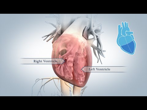
The human heart is a muscular organ that’s roughly the size of your fist. It has four main chambers:
- Right Atrium: This chamber receives deoxygenated blood from the body through veins called the superior and inferior vena cava.
- Right Ventricle: It pushes that blood to the lungs where it gets oxygenated.
- Left Atrium: After picking up oxygen, the blood returns to this chamber from the lungs via the pulmonary veins.
- Left Ventricle: This is where the magic happens—it pumps oxygen-rich blood out to your body through the aorta.
Each part plays a vital role in ensuring that your body gets what it needs to function well.
The Heart Valves: Checkpoints for Blood Flow
Now, let’s talk about heart valves. They’re like bouncers at a club—making sure only certain blood gets through at certain times. There are four main valves:
- Atrioventricular Valves: These include the tricuspid valve (between right atrium and right ventricle) and mitral valve (between left atrium and left ventricle). They keep blood from flowing backward when ventricles contract.
- Semi-lunar Valves: You’ve got the pulmonary valve (leading to lungs) and aortic valve (leading to body). They stop backflow when ventricles relax.
Think of them as gatekeepers, making sure everything flows smoothly!
The Electrical System: Keeping Beat
Ever wondered how your heart knows when to beat? That’s all thanks to its electrical conduction system. It works like this:
- Sinoatrial Node (SA Node): Known as the heart’s natural pacemaker, it creates electrical impulses that start each heartbeat.
- Atrioventricular Node (AV Node): This node acts as a relay station; it slows down those impulses before they travel to ventricles.
- Bundles of His & Purkinje Fibers: These carry signals quickly throughout ventricles so they contract effectively.
This system helps maintain a steady rhythm—like an orchestra playing in sync.
The Heart Wall: More than Just Layers
The heart has three layers:
- Epicardium: The outer layer that provides protection and houses coronary vessels.
- Myocardium: The muscle layer responsible for pumping action. It’s really thick on the left side since it has more work to do!
- endocardium:This innermost layer lines chambers and valves, keeping everything smooth for blood flow.
These layers all work together seamlessly. Each one plays an important role in keeping your ticker ticking!
Anecdote Time!
I remember being in a biology class when we had our first look at an actual heart. The teacher brought in one from a pig—it was surreal and kinda gross but also fascinating! We could see all those chambers and valves up close. Honestly, seeing how everything was laid out made me appreciate just how intricate our bodies are.
So there you have it—a straightforward look at cardiac anatomy! Understanding these structures can really enhance how you think about health. Your heart doesn’t just pump; it dances its own rhythm, keeping life flowing smoothly!
Detailed Heart Structure Diagram: Understanding the Anatomy and Function in Human Biology
So, let’s chat about the heart, shall we? It’s one of those organs that really steals the spotlight in our bodies. You know, like the lead singer in a band? The heart pumps blood all around, delivering oxygen and nutrients while also taking away waste. Pretty cool stuff!
Now, when you look at a detailed heart structure diagram, you’re going to see a few main features. First up are the four chambers—that’s like the different sections of your favorite room in your house! They include:
- Right atrium: This is where oxygen-poor blood enters from the body.
- Right ventricle: Blood then flows down here to get pumped to the lungs for some fresh oxygen.
- Left atrium: After picking up oxygen, blood returns to this chamber.
- Left ventricle: Finally, this is where the oxygen-rich blood gets pushed out to nourish your whole body.
But that’s just scratching the surface. Each chamber has its own job and works together like a well-oiled machine—sort of like how you and your friends coordinate for a group project!
The next big players are the valves. Think of these as little gates that make sure blood flows in only one direction. There are four main valves:
- Tricuspid valve: Separates the right atrium from the right ventricle.
- Pulmonary valve: Leads to the lungs for oxygenation.
- Aortic valve: Opens into the aorta, sending blood out to your body.
- Mitrial valve: Separates the left atrium from left ventricle.
These valves help prevent backflow, which is super important because nobody wants traffic jams in their bloodstream!
Now let’s talk about some fancy stuff: arteries and veins. Arteries carry blood away from your heart (like playful puppies eager to explore), while veins bring it back (kind of like them returning home after their adventure). The major artery leaving the heart is called the aorta, which branches out into smaller arteries.
Then we have something called blood circulation. There are two main loops going on:
– The **systemic circulation** sends oxygen-rich blood out through your body.
– The **pulmonary circulation** takes deoxygenated blood to your lungs.
Here’s where it gets kind of emotional! I remember my grandma telling me about her heart surgery. She had such courage! It was fascinating—and somewhat nerve-wracking—to hear how they replaced some valves and got her back on her feet. Her story reminded me just how vital this little pump really is.
Oh! And I can’t forget about **coronary arteries**. These guys supply blood directly to your heart muscle itself. Just think about it: Your heart needs its own food too! If these arteries get blocked, it can lead to something serious like a heart attack.
In sum, understanding the anatomy and function of our hearts helps us appreciate just how miraculous our bodies are. It’s not just about feeling that beat; it’s about all those tiny parts working harmoniously together day after day! So next time you spot a diagram of a heart, remember there’s so much more behind what makes it tick—and beat—and keep us alive!
You know, the heart is one of those organs that’s kind of taken for granted, right? We all know it pumps blood and keeps us alive. But when you really think about it, the complexities of how it works are pretty mind-blowing.
I remember the first time I saw a detailed heart diagram in my biology class. We had that old-school poster hanging on the wall, filled with labels and arrows pointing out everything from the atria to the ventricles. At first glance, it felt overwhelming—like trying to read a foreign language. But then slowly, as we dissected each part and understood its role, it was like watching a light turn on.
A comprehensive heart diagram isn’t just a pretty picture; it’s like a map to understanding our whole circulatory system. You’ve got these chambers working together—right atrium, right ventricle, left atrium, left ventricle—pumping oxygen-rich and oxygen-poor blood throughout our bodies. And then there’s all those valves ensuring blood flows in one direction. It’s almost poetic when you think about how well orchestrated everything is.
But here’s where things get really cool: visual learning! Seeing all these parts laid out helps you grasp things way better than just reading about them in a textbook. For example, when you understand that the right side of your heart handles deoxygenated blood coming from your body and sends it to your lungs for fresh oxygen—that makes so much more sense!
So yeah, if you’re ever feeling lost in science class or at home while studying, take a moment with a good heart diagram. Imagine what each part does as if they’re characters in a story—like the hero (the left ventricle) who sends oxygen-rich blood out to save the day! It’s fascinating how something that seems so complex can actually become clear with the right visual aid.
In short, diagrams can be warping but enlightening tools for scientific education. It connects dots between anatomy and function in ways our brains crave! Looking back on that poster from high school makes me appreciate how far we’ve come in understanding our own bodies—and hey, we owe it to diagrams like those for making learning way more engaging! So next time you see one of those comprehensive layouts of the heart or any other body part, take a moment to appreciate not just what they show but what they teach us about life itself!