You know what? The thorax kinda sounds like a mythical creature from some old tale, right? But it’s actually just a fancy term for the part of your body that holds your heart and lungs. Seriously, without it, we’d be in big trouble!
Picture this: You’re sitting at dinner, trying to explain why you can’t eat another bite because you feel stuffed, and your friend’s just staring at you like you’ve lost it. Well, that tight feeling in your chest? That’s your thoracic cavity doing its thing. It’s not just about food but about life—like breathing and keeping your ticker ticking!
So let’s take a moment to peek inside this incredible area of our bodies. It’s packed with cool structures and is way more intricate than you might think. You follow me? Buckle up; we’re going on a little tour through the human anatomy!
Comprehensive Guide to Thoracic Cavity Anatomy: Structure, Functions, and Clinical Significance in Human Biology
The thoracic cavity, you know, is that part of your body basically located above your abdomen and below your neck. It’s like a shield for some major organs. It’s fascinating how this space is built and functions, so let’s break it down!
Structure
The thoracic cavity has walls made up of bones and muscles. Here are some key features:
- Rib cage: This is like a protective cage around the organs. It consists of ribs, which are flexible but strong.
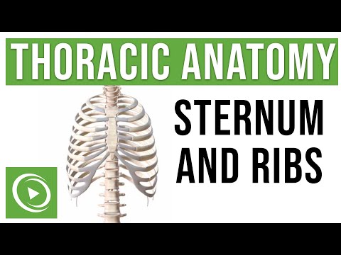
- Sternum: That’s the flat bone in the front, often called the breastbone. It connects with ribs to form a cage.
- Thoracic vertebrae: These are the bones in your spine that help keep everything stable while also allowing for movement.
- Diaphragm: This big muscle separates the thoracic cavity from the abdomen and plays a huge role in breathing.
Inside this cavity lies some pretty important stuff. You’ve got:
- Lungs: Obviously super important for breathing! They take in oxygen and expel carbon dioxide.
- Heart: This little engine pumps blood throughout your body—talk about essential!
- Mediastinum: A space between the lungs containing structures like the trachea and esophagus, plus blood vessels.
Functions
So what does all this do for you? Well, when you consider its main functions, they’re pretty crucial to keeping you alive:
- Your lungs work nonstop to bring fresh air into your bloodstream. Without that oxygen, you’re like a car running on empty!
- The heart manages blood flow, delivering nutrients and oxygen to every part of your body.
- The diaphragm helps move air in and out by contracting when you breathe—seriously cool muscle!
Clinical Significance
Now, let’s talk about why understanding this area is important from a medical point of view. Knowing the anatomy can help diagnose issues effectively!
- If someone has chest pain or difficulty breathing, healthcare providers need to figure out if there’s something wrong with their heart or lungs—could be anything from an infection to something more serious like pneumonia or even heart disease.
- If there’s damage or trauma to this area—for instance from an accident—it can threaten life quickly because so many essential structures are housed here.
I remember hearing a story about someone who survived a severe car accident largely due to their rib cage protecting their organs. They went through a tough recovery but were ultimately grateful their body held together as well as it did!
Anyway, that’s just a little peek inside the thoracic cavity! Its structure and function are vital both for everyday living and medical practice alike. Understanding its anatomy really highlights just how well everything works together in our bodies!
Comprehensive Thorax Anatomy Notes PDF for Medical and Biological Sciences
So, thorax anatomy, huh? It’s a super important area if you’re into medicine or any biological sciences. The thorax, or chest area, is like the body’s protective box for some of our biggest organs. Let’s break it down a bit.
First off, the thorax is bounded by the rib cage, which consists of 12 pairs of ribs. These ribs are essential because they protect vital organs like the heart and lungs. They’re not just decoration; they play a serious role in breathing too! When you inhale, your rib cage expands to let your lungs fill up with air.
Now about those organs. The lungs are two sponge-like things sitting pretty on either side of your heart. They have these tiny air sacs called alveoli, where all the gas exchange happens. Basically, oxygen comes in and carbon dioxide goes out. It’s like a little dance between gases!
Then there’s your heart—an incredible organ that pumps blood throughout your entire body. It’s tucked away behind the sternum, also known as the breastbone. The heart has four chambers: two atria and two ventricles, which work in harmony to circulate blood effectively.
And speaking of circulation, let’s not forget about the great vessels! You’ve got the aorta, which is like the main highway for blood leaving the heart, and then there are veins like the superior vena cava, bringing deoxygenated blood back from above.
Beyond organs, you’ll find muscles in this mix too! The major muscles include the intercostal muscles. They sit between each rib and help with breathing movements—contracting when we breathe in and relaxing when we breathe out.
In terms of nerves, that’s where it gets interesting! The thoracic spinal nerves emerge from your spine to control not just movement but also sensations throughout this area—pretty wild how everything connects.
So, if you’re thinking about comprehensive notes on thoracic anatomy for studies or just to get a better grasp on it all? Look for materials that cover these aspects thoroughly yet simply—the bones, muscles, organs, vessels, and nerves work together like a finely tuned machine.
That said: when discussing anatomy seriously, remember context matters! Detailed diagrams can be super helpful too—for visual learners especially!
In summary:
- The rib cage: protects vital organs.
- Lungs: site of gas exchange.
- The heart: pumps blood through chambers.
- Aorta & veins: manage blood flow.
- Intercostal muscles: assist in breathing.
- Nerves: regulate sensation & movement.
So there you go! A quick tour through thoracic anatomy without overcomplicating things too much. Just keep it simple: each part plays its role in keeping us alive and kicking!
Understanding Thoracic Wall Anatomy: A Comprehensive Guide to Structure and Function in Human Biology
So, let’s chat about the thoracic wall anatomy. You might be thinking, “What even is that?” Well, it’s basically the part of your body that protects your heart and lungs, and it has some pretty cool structures going on.
The thoracic wall is made up of a combination of bones, muscles, and connective tissues. You’ve got your rib cage, which is like a sturdy shield. It consists of **12 pairs of ribs** that curve around and connect to the spine at the back and to the breastbone (or sternum) at the front. The ribs are crucial because they not only keep your organs safe but also help with breathing.
Let’s break it down:
- Ribs: These guys come in two types: true ribs (the first seven pairs) connect directly to the sternum, while false ribs (the next five pairs) don’t have that direct connection.
- Sternum: This flat bone in the center of your chest connects with bones on both sides. Think of it like a bridge connecting vital structures.
- Costal cartilages: These are flexible tissues that attach ribs to the sternum. They allow for movement while still keeping everything in place.
Now, here’s where things get interesting: muscles! Yeah, they play a big role too. Your thoracic wall isn’t just about bones; it’s loaded with muscles that help you breathe.
The key muscle players include:
- Intercostal muscles: Located between each rib, these guys contract to help expand and shrink your chest cavity as you breathe in and out.
- Diaphragm: This dome-shaped muscle sits right below your lungs and is probably one of the hardest workers in your body—it does most of the heavy lifting when you inhale!
It’s wild to think how all these components work together seamlessly! Your rib cage expands when you take a deep breath—thanks to those intercostal muscles—and then snaps back into place when you exhale.
So picture this: you’re out running or playing basketball with friends on a sunny day. You start feeling winded after sprinting up that long hill—you know what I mean? That feeling happens because your thoracic wall is working hard! Your diaphragm pulls downwards creating more space for air to fill your lungs. It’s pretty amazing how all this happens automatically without you even having to think about it.
And let’s not forget about blood vessels and nerves! The thoracic wall has major blood vessels running along side—like arteries—that supply oxygenated blood to tissues around your chest area. Plus there are nerves too—like intercostal nerves—telling those muscles when to move 💪.
If something goes wrong in this region—like injury or disease—it can affect breathing or cause pain which makes it super important for health professionals to understand this structure well.
So now you know how cool—and complex—the thoracic wall really is! It’s an essential part of our anatomy that protects vital organs while helping us take those much-needed breaths throughout our lives. And honestly? It’s a great reminder of just how wonderfully made our bodies are!
Thoracic anatomy is like, this fascinating mix of complexity and elegance. Think about it: your chest is home to some of the most vital organs, not just your heart and lungs, but also all those intricately connected structures that keep you alive and kicking.
I remember a buddy of mine, totally into sports, who had a nasty fall while playing basketball. He thought he just knocked the wind outta himself. But, after getting checked out, it turned out he had cracked a rib. I’ll never forget how serious that moment became when the doc explained how even a small injury in that area could mess with breathing or put pressure on his lungs. It struck me then just how delicate and essential this whole thoracic region is.
So, what exactly’s going on in there? Your rib cage protects everything inside while allowing for the necessary movement during breathing. The sternum—or breastbone, as some folks call it—plays a huge role too; it connects with ribs to help form this sturdy shell around your chest cavity. And those lungs? They’re like this amazing sponge system absorbing oxygen and getting rid of carbon dioxide. It’s wild to think about how much happens with every breath you take.
And let’s not forget about blood vessels! The thorax houses major arteries and veins—like the aorta—transporting oxygenated blood all over your body. It’s like this intricate highway system running beneath our skin. And if you were to peek inside during an operation (not advising you to do that!), you’d see how everything’s so interconnected.
What gets me though is the way our bodies work in harmony most times without us even realizing it! You go through life breathing without thinking about each expansion of your rib cage or those tiny little movements in your diaphragm that make it all happen.
In short, looking at thoracic anatomy gives us insight into something so critical yet often taken for granted. It’s pretty humbling to think about how fragile we are underneath all that tough exterior and muscle. Each breath is more than just air; it carries life itself—and understanding that can really change how you view everything going on inside ya!